 |
||
Home > TERATEC FORUM > Workshops
Tuesday June 22 - Application Workshop
Workshop 02 - 16:00 to 18:00
Communicable diseases and vision disorders
Chaired Philippe Gesnouin, Tech Transfer Associate for Life Science and Healthcare, Inria and Richard Bosmans, Directeur des Partenariats et Financements Externes de la R&D, Essilor International
Computational imaging of the eye with near infrared light by digital holography
By Michael Atlan, Researcher, French National Center for Scientific Research (CNRS)
|
ye conditions are major global health problems. The number of people living with blindness and visual impairment is expected to reach 100 million and 600 million respectively by 2050. The most frequent pathologies: macular degeneration, diabetic retinopathy, glaucoma and vascular occlusion, are accompanied by structural and functional alterations of local microvascular networks. For these diseases, state-of-the-art methods for blood flow analysis are lacking precision and are not compatible with patient monitoring. The establishment of new methods for the reliable measurement of hemodynamic contrasts in the eye would facilitate the management of the disease in these patients.
One particular technique involving coherent near-infrared light detection may accelerate the transition to computational imaging in ophthalmology, and open the way to non-invasive measurements of paramount importance. With the exponential availability of computer resources and advances in ultra-fast cameras, the next revolution in eye imaging will likely be fueled by imaging approaches derived from digital holography and laser radar techniques that enable optical wavefield measurements and their digital manipulation from rapid recordings of optical interferograms.
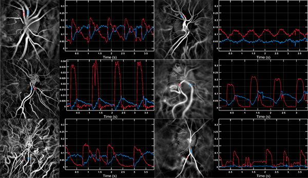
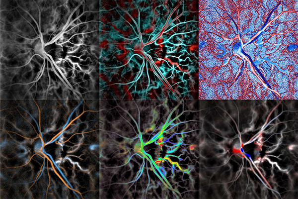
In that context, we developed and used an innovative medical holographic imaging tool for ophthalmology which reveals quantitative biomarkers with millisecond temporal resolution. This instrument enables simultaneous quantitative measurements of hemodynamic parameters in the choroid, retina, iris and conjunctiva, including discrimination of arteries and veins, and measurement of pulse waves and spectrograms in retinal vessels, directional blood flow in the lumen of the central retinal artery and vein. Holographic imaging also enables assessment of the transparency of the anterior segment, the dynamics of the refractive state, and comprehensive 3d eye movements with millisecond resolution.
 LDH in a variety of eyes. From left to right and top to bottom: Healthy eye, Takayasu's disease, central retinal vein occlusion x2, arrhythmia, glaucoma. |
 |
Biography: Michael Atlan is a researcher at the French National Center for Scientific Research (CNRS) in experimental optical physics, instrumentation, and computational imaging. He works at the Langevin Institute (CNRS UMR 7587), and the Quinze-Vingts National Eye hospital (INSERM CIC 1423) in Paris. He develops coherent-light instrumentation and numerical processing methods for laser Doppler vibrometry, microscopy, and blood flow imaging in low-light. His expertise is focused on methodologies involving optically-acquired digital holograms for sensitive coherent imaging at high throughput. He is the project manager of the versatile real-time digital hologram rendering software "Holovibes" (http://holovibes.com), and the co-author of journal articles, patents, and book chapters on the subject of holography. |
|---|
Register now and get your badge here
- TERATEC Forum is strictly reserved for professionals.
- Participation to exhibition, conferences and workshops is free (subject to seats available)
- On line registration is obligatory.
